
DEPARTMENT
KERATOCONUS CURE
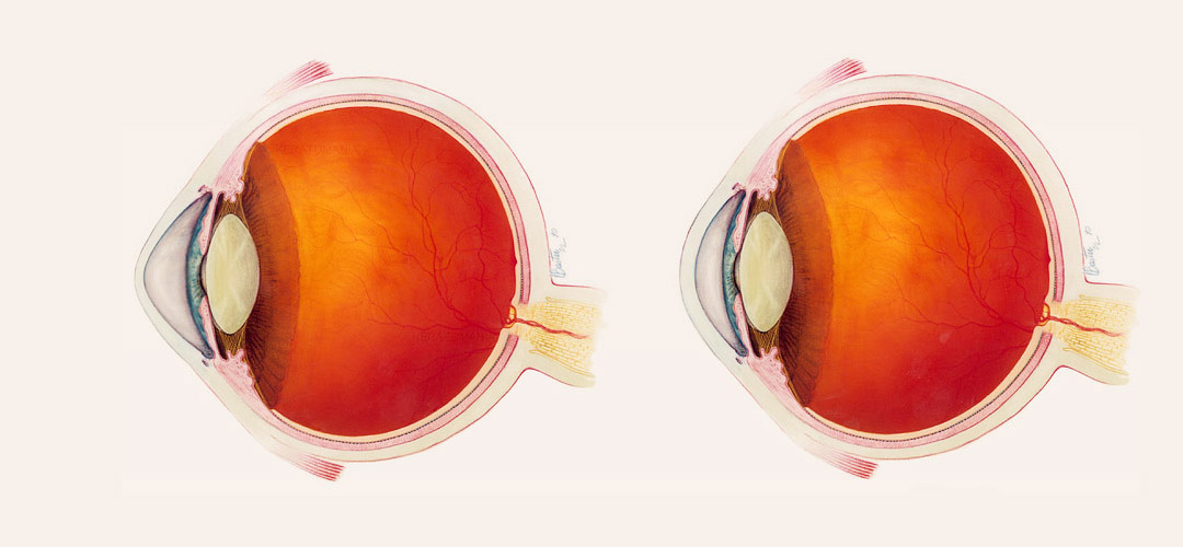
Dystrophic (associated with impaired tissue supply), degenerative changes in the cornea of the eye in some cases lead to a pronounced deformation of this external transparent protective layer. The cornea extends forward, acquiring a peculiar, pointed, conical shape. Accordingly, the refractive characteristics change roughly: refraction of light in this form excludes a clear distinction of objects.
This condition is called “keratoconus” and is a serious ophthalmologic pathology requiring timely qualified intervention. The sharpness of vision decreases rapidly, various optical aberrations occur (distortion of the shape of visible objects, halo effect around light sources, “rainbow” blurriness of contours, doubling or multiple duplication of objects), the ability to binocular vision is lost; in some cases, pain syndrome develops, the corneal membrane becomes turbid. A thorough examination is necessary, including precise topological measurements of the geometry of the keratoconus, biomicroscopy, tomography, ophthalmometry, etc. Reverse development of this state does not detect, and to eliminate it resorted to one or another – depending on the specific clinical picture.
Keratoconus, fortunately, refers to relatively rare diseases: in the total volume of ophthalmologic pathology its proportion ranges from 0.01% to 0.6% of cases. It should be emphasized, however, that these estimates are approximate and ambiguous, since a number of factors affect the incidence rate, for example, geographical or racial (there are statistical data that the Mongoloids fall ill more often). There is no consensus about whether the risk of developing keratoconus depends on sex. Medico-statistical studies are complicated, in particular, by the difficulties of differential diagnostics in the early stages – for example, between the initial development of keratoconus and complex astigmatism.
It is definitely established that the “start” of keratoconus deformation in most cases occurs at pubertal age, during a period of turbulent puberty. Almost always (95% of cases) affects both eyes. With regard to the nature of the development of the disease (possible variants – slow growth, spasmodic flow with periods of temporary improvement, spontaneous arrest, rapid progression of pathology, acute keratoconus), it is highly individual and varies even on the right and left eye of the same patient.
Causes of keratoconus
Due to a number of circumstances (rare occurrence, complexity of diagnosis, etc.), keratoconus has not been sufficiently studied to date. First of all, it concerns the causes and etiopathogenetic mechanisms of the disease. The scientific literature reports on several factors that can be triggered with varying degrees of reliability: genetic, endocrine, metabolic, immune, etc. The most reasoned is the polyethological hypothesis, according to which conical deformation of the cornea is due to hereditary predisposition in combination with metabolic and immune disorders; this combination is especially dangerous precisely during periods of intensive and rapid hormonal adjustment (which, in particular, is observed in adolescents). Various sources also reported on the possible impact of the environmental factor (radiation background, air pollution), excessive insolation (exposure to the ultraviolet part of the solar spectrum), there were speculations about the provocative role of various diseases (allergic, infectious-inflammatory, traumatic, genetic). Finally, recently, alarming reports have appeared of frequent cases of keratoconus due to post-operative keratotectasia, which in turn sometimes becomes a side effect of laser vision correction.
It is known that cardiac biochemical changes occur in the tissue of keratoconus affected by the keratoconus: the concentration of collagen and protein decreases, free radicals are formed, etc. As a result, the elasticity of the corneal layer decreases sharply, it stays in constant rigid tension and gradually extends into the cone.
Classification of keratoconus
Depending on the context (theoretical, research, diagnostic, therapeutic), different approaches and criteria for classification of diseases are used in medicine. So, genesis keratoconus is divided into primary and secondary. “Secondary” means a known, established cause of development – and most cases of secondary keratoconus are caused, unfortunately, by iatrogenic causes, i.e. medical intervention.
Several different classifications were proposed for the stages of development; among them the most widely spread version was the Swiss ophthalmologist Mark Amsler, consisting of four gradations.
Keratoconus in the first stage is manifested by astigmatism (inability to clearly focus) and a slight decrease in visual acuity, which can be optically corrected – cylindrical glasses or lenses. In the second stage, astigmatism also lends itself to optical correction, but visual acuity is reduced to 0.4-0.1 of the norm. In the third stage of keratoconus, the processes of thinning and stretching the cornea anteriorly are intense; vision deteriorates to 0.1-0.02, which can be corrected only by rigid contact lenses. Finally, the fourth stage is characterized by pronounced conical deformation, a decrease in visual acuity in a non-correctable degree (0.02-0.01), turbidity of the corneal membrane.
Symptoms of keratoconus
The clinical picture of the keratoconus is determined mainly by optico-geometric patterns: as the cone of the cornea increases, – the transparent refractive medium – the focal point shifts forward from the retina (myopia progresses). Almost immediately, astigmatic symptoms (mismatch of the refraction axes, general defocusing) are joined, the indices of which constantly change in dynamics. A typical symptom is also monocular diplopia, the bifurcation of the objects observed by the diseased eye, and soon the number of illusory “copies” increases significantly.
The instability of refractive disorders forces the patient to change glasses frequently, but optical correction, even calculated by an ophthalmologist with high accuracy, gives only an incomplete and short-term effect: in cases of rapid disease progress, the situation sometimes changes after the receipt of a prescription for new glasses and their manufacture. Soft contact lenses are also becoming less effective, since their dense adherence to the cornea does not correct from a certain moment, but merely repeats the anomalous curvature of the surface.
Many patients complain about linearity distortions (straight lines are seen curved, the letters in the printed text lose their habitual form, which makes reading extremely difficult), note halo-halos around the luminous objects, painfully sharp sensitivity to light, carvings, burning, itching, “sand” in eyes. At later stages, the phenomenon of “night blindness” (sharp drop in vision in the twilight or twilight) is replaced by a decrease in light sensitivity as such. Meanwhile, the characteristic protrusion of the cornea gradually becomes noticeable and obvious.
Despite the wide range of statistical indicators, in general keratoconus is considered to be a slowly progressing disease. From the beginning of the process to the formation of the developed symptomatology inherent in the fourth stage, it usually takes 10-15 years; approximately half of the patients develop keratoconus for a long time, it stops or drastically slows down in the first stage. However, 5-10% of patients rapidly develop the so-called. acute keratoconus: Descemet’s membrane is torn (posterior border membrane), the corneal substance is filled with fluid, which is accompanied by intense pain syndrome and corneal edema. The acute phase of this condition lasts up to 3 weeks, during which the symptomatology is gradually reduced (as the corneal tissue becomes scarred). Sometimes the curvature of the cornea after an acute keratoconus somewhat normalizes, approaching a spherical.
Treatment of keratoconus
Depending on the stage, peculiarities of the course and rates of keratoconus development, both conservative treatment and ophthalmosurgical intervention can be indicated.
Conservative therapy includes, first of all, optical correction. In the early stages, – with stabilization of the process and a slight decrease in vision, – appoint glasses; with more severe symptoms, contact lenses are shown. For cases where soft lenses no longer cope with their task, in recent years, manufacturers have offered combined “semi-rigid” lenses (with a soft perimeter and a rigid central part that mechanically presses the protruding region of the cornea). Vitamin and antioxidant complexes, immunomodulators and stimulants, preparations for activation of blood circulation and tissue nutrition, physiotherapy (phonophoresis, magnetotherapy, etc.) are prescribed to improve the general condition of the visual system and prevent further progression of the pathological process.
An effective method of therapy of keratoconus is the removal of the superficial epithelial corneal layer, followed by instillation of riboflavin and ultraviolet irradiation. This method, called “cross-linking,” serves to strengthen the cornea and increase its elasticity, i.e. resistance to stretching. In some cases, cross-linking manages to suspend the process or even wrap it back – by softening the already formed refractive anomaly – and then returning to normal optical correction (glasses, soft contact lenses). At the first stages of keratoconus development, laser correction (photorefractive and phototherapeutic keratectomy techniques) can be shown and appropriate: the myopia and astigmatic component are eliminated, the outer layer of the corneal membrane is strengthened.
In other cases, thermokeratoplasty can be prescribed – the coagulant thickens those areas of the corneal shell that “lag” behind the central apex of the cone protruding forward. Other surgical options include the implantation of stromal rings (stroma – the main corneal tissue) or the donor cornea as a whole. The latter option, i.e. classical through keratoplasty, can be applied even at the final stages of keratoconus deformation; This method has a very high probability (up to 90-95%) of the chance of engraftment of the donor material and effectiveness in terms of increasing visual acuity.In cases where thermokeratoplasty can be prescribed, those zones are thickened by the coagulantcorneal shells that “lag” behind the central apex of the cone projecting forward. Other surgical options include the implantation of stromal rings (stroma – the main corneal tissue) or the donor cornea as a whole. The latter option, i.e. classical through keratoplasty, can be applied even at the final stages of keratoconus deformation; This method has a very high probability (up to 90-95%) of the chance of engraftment of the donor material and effectiveness in terms of increasing visual acuity.
BOOK ONLINE
Address
Qemal Stafa Str, Apt 31, 3-rd C Floor
Near the General Prosecutor’s Office
klinika.uzllova@yahoo.com
info@eye.al
Phone
0682074039
04 22 52 306

